Best Guard Dogs for First Time Owners & Families With Kids
 Additional expenses can differ significantly between clinics, so it’s extremely troublesome to say how a lot it could be. The cost of your dog’s microchip will differ based mostly on where it’s accomplished. Shelters and rescues typically have the sources to place microchips into their pets before they're adopted. Some of those facilities will charge you an additional charge to microchip your dog, however you probably can usually count on it to cost $20–$50. The accidents-only cowl is for the costs in case your canine unexpectedly gets hurt and needs emergency care. It’s just a fundamental cover that doesn’t include regular vet checks or routine care.
Some signs of heart disease in pets are:
Si la fuga está localizada en un hemitórax, va a ser preciso realizar una toracotomía del costado, ya que da el más destacable abordaje a las construcciones de tal lado. Si la afección es bilateral o no se identifica el sitio exacto de la fuga, se debe realizar una toracotomía exploratoria por esternotomía media 7. Asimismo hay que considerar que en los traumatismos torácicos cerrados es recurrente la presencia de contusiones pulmonares (Figura 5) y sangrados mediastínicos concomitantes o no con el neumotórax. La toracocentesis bilateral es un trámite tanto diagnóstico como terapéutico de neumotórax y esta se debe realizar antes de tomar las radiografías después de un traumatismo torácico.
Radiografía para perros: ¿Cuánto cuesta en México en promedio?
Además, con los sistemas de radiografía digital, una cantidad excesiva de exposición fuera del sujeto puede ofrecer lugar a una falsa interpretación de los datos por la parte del algoritmo de reconstrucción y degradar substancialmente la calidad de la imagen. Si esto sucede, la exposición debe repetirse con una colimación correcta para lograr una imagen aceptable. En la mayoría de los casos, el haz de rayos X debe colimarse a ~1 cm fuera de los límites del sujeto para otorgar una calidad de imagen óptima y protección radiológica para el plantel. La colimación adecuada del haz de rayos X no puede sustituirse por la utilización de la herramienta de recorte de imágenes disponible en la mayor parte de los sistemas de software utilizados para generar imágenes digitales. Esta es una herramienta de posprocesamiento y no afecta a la calidad de la imagen ni a la reconstrucción. Además laboratorio de exames animais esto, esta herramienta jamás debe usarse para recortar ninguna anatomía del paciente capturada por la exposición inicial y la reconstrucción. El posicionamiento apropiado también es esencial para maximizar el contenido diagnóstico del examen de rayos X.
¿Cuánto cuesta hacer una resonancia a un perro?
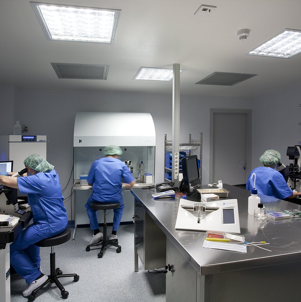
Este procedimiento nos permite advertir quézona cardiaca concreta está aumentada de tamaño(fig. 6). El autorXavier Sánchez SalgueroDoctor en Veterinaria por la Universidad Autónoma de Barcelona (UAB) en 2012. En la actualidad es copropietario y veterinario especialista en diagnóstico por imagen en la Clínica Veterinària 4 Vents de Blanes(Girona). Asimismo es coordinador de cursos en línea de veterinaria en Cursovet.Durante los cuatro años de máster y tesis doctoral fue docente de radiología en la Facultad de Veterinaria de la UAB realizando ecocardiografías, estudios radiográficos y ecografía abdominal.
Fat infiltration in the cranial mediastinum can mimic a mass (cats and dogs). This elevated width normally has parallel sides, as seen on the VD/DV view, not like an enlarged lymph node or thymoma. In the middle mediastinum the fat adjacent to the guts may silhouette with the cardiac outline mimicking heart enlargement. Caudal mediastinal widening, between the accent and caudal left lung lobes may be mistaken for pleural effusion. Infestation by the heartworm Dirofilaria immitis happens in each canine and cats.
This could be completed by utilizing excessive kVp, low mAs exposure settings. The high kVp setting decreases distinction, Demotest.Ca and offers added latitude and room for technical error (Digital radiology systems accomplish this same thing). A good technique will permit faint visualization of the spine via the cardiac shadow on ventrodorsal (VD) or dorsoventral (DV) views. As for another physique half, a grid is important if the thorax is thicker than 10 cm. Small canines and non-obese cats measuring lower than 10 cm may be imaged using table top (non-grid) approach and a detail (slow film-screen combination) cassette.